Why Do Some Anterior Implant Restorations Look More Natural?
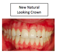
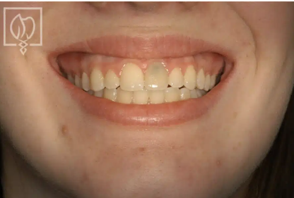
Anterior implant restoration with natural-looking crown
The hardest tooth in the mouth to restore is one of the four maxillary incisors, because they are directly in the line of sight and are being compared to the other incisors next to them. Even more difficult than that is the restoration of an implant in that location. Unless the implant is precisely placed using a very accurate surgical guide, the restoration can end up looking bulky, too opaque, and not a match for the adjacent teeth. The difference between a stunning restoration and a disappointing one often comes down to millimeters of implant positioning.
The Importance of Precision Surgical Planning
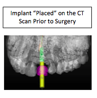
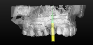
At Prosthetic Dentistry of Washington, D.C. (located in the Washington, D.C.-Bethesda area), Dr. Marlin uses “guided surgery” using custom surgical stents when inserting all of his implants. Using extensive presurgical planning via a CT Scan (3D image) of your jaw, Dr. Marlin is able to precisely plan the angle, depth, and location of each implant prior to the surgery as he “places” them directly on the CT Scan computer model. In many cases, he has the CT Scan software company create the surgical guide, especially when inserting anterior implants where the aesthetics has to be very exact.
This precision planning approach represents a significant advancement over traditional freehand implant placement. By using the 3D data to evaluate your bone anatomy, sinus position, nerve channels, and adjacent tooth positions, Dr. Marlin can determine the optimal placement for your implant before he ever picks up a surgical instrument. This reduces complications, increases implant success rates, and ensures that the final restoration will look natural and integrate seamlessly with your smile.
Ideal emergence profile for anterior implant restoration
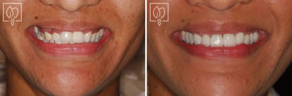
Final anterior implant restoration integrated seamlessly with adjacent teeth
Designing the Emergence Profile
The precise planning allows him to actually design how the abutment that comes out of the implant and holds the crown will be located in relation to the restoration. This is crucial for anterior teeth because the emergence profile, the contour where the crown emerges from the gum tissue, is visible to anyone looking at your smile. If the implant is placed too far facially, the crown will look bulky. If it’s placed too far lingually, the crown will look too transparent or thin. The ideal position allows the ceramist to create a crown with natural contours, proper thickness, and translucency that perfectly mimics the tooth being replaced.
How It Works: The Path to Natural Results
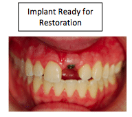
After four to six months, the implant has integrated to the bone and is ready for restoration. The process is usually very predictable, since the implant (the replacement tooth root) has been precisely placed according to the preoperative plan. A custom abutment is fabricated to create a very good-looking crown that has a natural emergence profile and integrates beautifully with the gum tissue.
The collaboration between surgical precision and prosthodontic artistry is what transforms an implant from a functional replacement into a restoration that is indistinguishable from natural teeth, combining the benefits of implant dentistry with masterful crown artistry. Dr. Marlin’s commitment to precision surgical placement combined with our in-house laboratory expertise ensures that your anterior implant restoration will be both functionally superior and esthetically superior to the original tooth.
The Result: a happy patient!
If you’re considering anterior dental implants and want the most natural-looking result possible, working with a specialist who invests in precise surgical planning and has access to an in-house laboratory makes all the difference.
Related Patient Success Stories
Explore similar patient success stories demonstrating our expertise in advanced prosthetic dentistry.

Temporary Crowns Restore Patient's Smile in Just One Day with an Immediate Smile Makeover
A patient from Potomac, Maryland, came to Elite Prosthetic Dentistry with the chief complaint of pain from a failing dental implant and its significant impact on her appearance.
Multi-Faceted Treatment for Patient Unhappy With Her Artificial-Looking Crowns, Teeth and Gums
Many patients come to Elite Prosthetic Dentistry unhappy with the appearance of their smile. However, this particular patient presented with multiple interconnected problems that together created a smile she found deeply unsatisfying.

Treating Kevin’s Collapsed Bite with a Complete Smile Makeover with New Dentures
Dentures are sometimes not created to the ideal aesthetic and functional scheme. When improperly fabricated, dentures can make an individual appear almost a generation older than their actual age. They can have a poor fit that feels loose and unstable when eating or speaking, and they can actually accelerate bone loss over time.
Related Articles
Deepen your knowledge with additional insights on this topic.
 Dental Implants
 If a Single Front Tooth is Replaced with an Implant, can it Look Natural?
A single front tooth implant can look completely natural with precision placement, custom abutments, and hand-crafted porcelain crowns. Washington, DC.
 Dental Implants
 What is Precision Implant Placement (PIP)?
Learn what Precision Implant Placement (PIP) is and how meticulous planning ensures optimal implant positioning for long-lasting results in Washington, DC.
 Dental Implants
 What is the ideal Surgical Guide for Precision Implant Placement?
CBCT-based surgical guides allow virtual implant planning for precise positioning in optimal bone, ensuring predictable results in Washington, DC.