What Causes Dental Implant Failure, and How Frequent Is It?
Dental implant failure is usually not a sudden event. It is a process, and it almost always follows the same path: the gum tissue around an implant becomes inflamed (a condition called peri-implant mucositis), the inflammation advances into the supporting bone (peri-implantitis), and if nothing interrupts it, the implant is eventually lost. Understanding that sequence is the key to both preventing failure and repairing a failing implant in time.
How frequent is it? The honest numbers deserve to be quoted. A systematic review and meta-analysis by Atieh and colleagues, covering 1,497 participants, found peri-implant mucositis in 63.4% of participants and peri-implantitis, the actual bone loss, in 18.8% (1). Implant dentistry as a whole has a problem it does not always advertise.
Our own experience is different: more than 97% of our patients still have a healthy implant after 20+ years. That is not luck, and it is worth explaining why.
Three Causes That Are Built In Before the Implant Ever Fails
When implants develop bone loss, the roots of the problem are frequently laid down on day one. Three failure paths dominate (2,3): first, incomplete planning that does not fully account for bone architecture and the ideal location of the implant together with its future restoration; second, placing implants freehand, without a surgical guide, resulting in poor positioning; and third, abutments and crowns fabricated with an inadequate emergence profile, the contour where the restoration meets the gum, so the site traps plaque and resists cleaning.
Notice what these have in common. None of them is the patient’s fault, and all of them are preventable with disciplined technique.
What Poor Positioning Looks Like
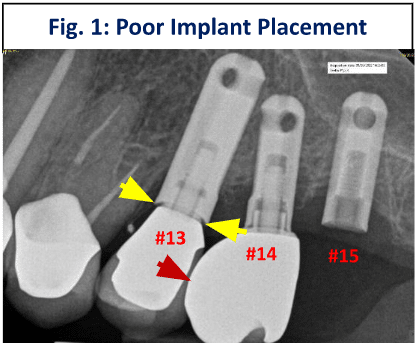
Fig. 1: Poor Implant Placement
In Fig. 1, implants #13 and #14 sit too close together because they were not placed using a surgical guide generated from virtual planning. As a consequence, the abutment on #13 could not seat completely, leaving a gap (yellow arrows) that harbors bacteria and invites peri-implantitis. The crown on #14 is over-contoured, making it plaque-retentive and difficult to clean, a setup for gum inflammation. Implant #15 was not restorable at all.
One radiograph, three separate seeds of failure, all planted at placement.
How Ideal Positioning Is Engineered
Every implant case in our practice starts with a CBCT scan to measure bone width, angulation, and density. That analysis determines whether the site first needs bone grafting or ridge augmentation, and the implant is then placed virtually, in its ideal position, on the scan itself.
From that virtual plan we fabricate a precise surgical guide that transfers the planned position exactly into surgery (2). We also plan for healthy gum tissue around the implant, because soft tissue is part of the defense. And when the implant is ready to restore, our in-house laboratory technician creates the natural emergence profile that keeps the site cleanable, with no compromises forced by a misplaced implant.
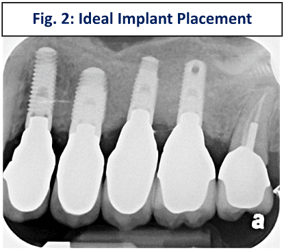
Fig. 2: Ideal Implant Placement
In Fig. 2, the implants were seated with a CT-generated surgical guide, allowing crowns with proper emergence profiles to be created easily. This is what boring, uneventful implant longevity looks like on a radiograph.
The Patient’s Side of the Ledger
Planning and placement set the ceiling, but habits and health still matter: hygiene, smoking, uncontrolled diabetes, and heavy grinding forces all influence outcomes. We cover those in detail in common factors that can limit implant success, and the mechanism of bone loss itself in what causes failed implants due to bone loss.
If You Suspect Your Implant Is Failing
Bleeding or puffy gums around an implant, a bad taste, looseness, or bone shadowing on an X-ray all justify prompt evaluation, because an implant caught early is often saveable. Dr. Gerald Marlin is a specialty-trained prosthodontist with more than 3,900 implants placed and restored, and evaluating troubled implants is a core part of our practice. Call 202-244-2101 or request a consultation at Elite Prosthetic Dentistry in Friendship Heights, Washington, DC.
Sources
- Atieh MA, Alsabeeha NH, Faggion CM Jr, Duncan WJ. The frequency of peri-implant diseases: a systematic review and meta-analysis. J Periodontol. 2013 Nov;84(11):1586-98. doi: 10.1902/jop.2012.120592. Epub 2012 Dec 13. PMID: 23237585.
- Abdelhay N, Prasad S, Gibson MP. Failure rates associated with guided versus non-guided dental implant placement: a systematic review and meta-analysis. BDJ Open. 2021 Aug 18;7(1):31. doi: 10.1038/s41405-021-00086-1. PMID: 34408127; PMCID: PMC8373900.
- Gomez-Meda R, Esquivel J, Blatz MB. The esthetic biological contour concept for implant restoration emergence profile design. J Esthet Restor Dent. 2021 Jan;33(1):173-184. doi: 10.1111/jerd.12714. Epub 2021 Jan 20. PMID: 33470498.
See How We Resolve These Problems
Our patient success stories show real cases and real results. Browse outcomes from a specialist prosthodontist with decades of experience and 3,900+ implants placed.
Key Takeaways
- ✓ Implant failure is usually a disease process, not an event: gum inflammation (peri-implant mucositis) progresses to bone loss (peri-implantitis), which can ultimately cost the implant.
- ✓ The published numbers are sobering: one large meta-analysis found peri-implant mucositis in 63.4% of participants and peri-implantitis in 18.8%.
- ✓ Three preventable causes drive much of it: incomplete planning, placement without a surgical guide, and crowns or abutments with a poor emergence profile.
- ✓ Careful CBCT-based planning, guided placement, and in-house restoration design are how those causes are engineered out of treatment.
- ✓ A failing implant caught early is often salvageable, which makes evaluation urgent rather than optional.
Frequently Asked Questions
How common is dental implant failure?
Outright loss of an implant is relatively uncommon, but the disease that leads to it is not. A systematic review and meta-analysis covering 1,497 participants found gum inflammation around implants (peri-implant mucositis) in 63.4% of participants and actual bone loss (peri-implantitis) in 18.8%. Those conditions, left untreated, are how implants eventually fail.
What is the difference between peri-implant mucositis and peri-implantitis?
Mucositis is inflammation and infection of the gum tissue around an implant, and it is reversible when treated. Peri-implantitis is the next stage, where the infection begins destroying the bone that supports the implant. Bone loss is far harder to undo, which is why early treatment of gum inflammation matters so much.
What actually causes implants to fail?
The most preventable causes are built in before the implant ever carries a tooth: incomplete planning that ignores bone architecture, freehand placement without a surgical guide, and abutments or crowns contoured so poorly that they trap plaque or leave gaps that harbor bacteria. Patient factors such as hygiene, smoking, and heavy bite forces then compound the risk.
Can a failing dental implant be saved?
Often, yes, especially when bone loss is caught early. Depending on the cause, treatment can include decontaminating the implant surface, treating the gum infection, regrafting lost bone, or redesigning a faulty crown. The longer the disease progresses, the fewer options remain, so evaluation should not wait.
Related Patient Success Stories
Explore similar patient success stories demonstrating our expertise in advanced prosthetic dentistry.
 Before
  After
After How a Front Tooth Lost to Childhood Trauma Was Rebuilt with Bone Grafting and a Long-Lasting Implant
A teenager was referred by her father after earlier trauma left her upper left front tooth slowly failing from root resorption. She was still growing, so an immediate implant was the wrong move. The tooth had to be maintained to buy time, then replaced correctly once she reached skeletal maturity.
 Before
  After
After Implant Supported Reconstruction: Failing Bridgework and Missing Back Teeth Rebuilt with Coordinated Specialist Care
Referred by another dental specialist with severe bone resorption on the upper left, multiple broken-down lower teeth requiring extraction, and failing lower back teeth that had left the bite without solid support. No single procedure, and no single provider working alone, could rebuild a situation this interconnected.
 Before
  After
After How a Loose Upper Bridge and Aging Crowns Were Rebuilt with Staged Implant Reconstruction
A patient referred by her general dentist after years of aging dentistry no longer holding up. A loose upper bridge and crowns more than twenty years old, combined with the effects of advanced periodontal disease and severely compromised tooth abutments, required a staged surgical and restorative plan delivered with comfort planning at the same time.
Related Articles
Deepen your knowledge with additional insights on this topic.
Bone Grafting & Surgical Rebuilding a Ruined Implant Site with Severely Damaged Bone: The Three-Stage Protocol
Debridement, growth-factor grafting, CBCT-guided planning: the step-by-step protocol that turns a severely damaged implant site into a healthy ridge.
 Bone Grafting & Surgical
 What Is Ridge Augmentation Bone Grafting?
Ridge augmentation rebuilds a jaw ridge too narrow or angled for an implant. See real measurements from planning to precise placement. Washington DC.
 Bone Grafting & Surgical
 What Is a Partial (Internal) Sinus Lift?
A partial (internal) sinus lift adds bone at a single implant site through the implant preparation itself, often with the implant placed the same visit.